不想错过界哥的推送?
戳上方蓝字“
医学界影像诊断与介入频道
”关注我们
并点击右上角“···”菜单,选择“
设为星标
”

曾经,一看到肺动脉血管成像(CTA)上有充盈缺损,就大呼:
肺栓塞了,肺上长血栓了!
立马抗凝、对症治疗。
后来发现,
肺动脉造影才是金标准
。我就纳闷:肺动脉CTA上发现了充盈缺损,那不是血栓会是神马?
再后来,内科学第八版第103页:
肺动脉造影诊断肺栓塞的特异性是95%-98%
。我就更纳闷了,肺动脉造影作为金标准,为什么还会误诊肺栓塞?特异性居然不是100%。
再后来,我知道癌栓。但是除了手术活检,其他办法是不可能很准确地鉴别癌栓啊,还是得抗凝啊!
曾经有人问我:肺栓塞老是治不好,为什么?
我知道很多原因,但是我居然不知道一个很简单的原因:
肺栓塞治不好,有可能那根本就不是肺栓塞!
看了很多文献,才发现,自己曾经真的是too young,too simple。
年轻不知晓,缺损都栓了。
夜来看文献,误诊知多少。
为此,我来写写肺栓塞CTA葵花宝典残页,为什么叫残页呢?我只是分享一些经验,还木有水平写整本葵花宝典。
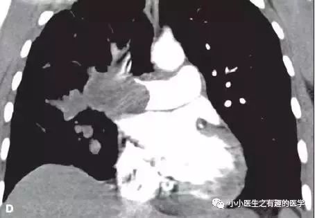
首先,来看看这个患者。美国康涅狄格州立大学报道,58岁男性,咳嗽,劳力性呼吸困难,进行性双侧下肢水肿2月。
A 58-year-old man presented with 2 months of cough, dyspnea on exertion, and progressive bilateral lower extremity edema.
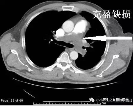
这是什么?妥妥的肺栓塞啊,还能有啥?
怎么办?肿么办?
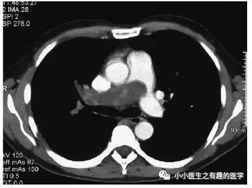
别慌,米国人民的治疗和我们一样。抗凝治疗,2周后,患者呼吸困难加重返院。
He was treated expectantly with anticoagulants for pulmonary embolism but returned 2 weeks later with worsening dyspnea and echocardiographic evidence of right heart failure.
丹雄OS:
在中国,很可能家属第一句话:医生,你给我老爸用的什么狗屁药,你个庸医,患者本来好好的,被你越治越重。上次来看病,本来没什么病的,我们来医院主要是旅游、参观的。
是的,有些人来医院并不是看病的,我们医生也没注意这个问题。有一次,科室的电脑活生生被人带出医院,神不知鬼不觉,监控录像发现了,医生还被扣了奖金,监控录像就是来扣奖金的,不是监视贼的,也抓不到贼。
抗凝治疗,病情加重,怎么办?这下该好好想想,肿么办?

这些医院都是很牛叉的。
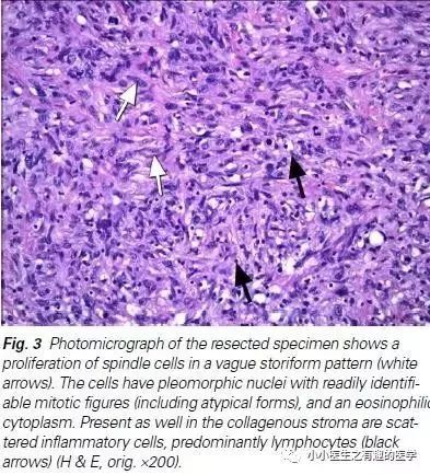
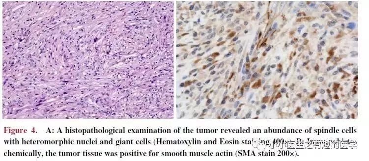
行动脉血栓内膜剥脱术,然而结果示黏液性梭形细胞肉瘤。
Surgical thromboendarterectomy revealed a high-grade myxoid spindle cell sarcoma.
是的,
这不是肺栓塞,这是肺动脉内膜肉瘤
。这不是扯蛋肺栓塞,这是扯蛋肺动脉肿瘤。扯蛋是肯定的,然而,血栓且不肯定。
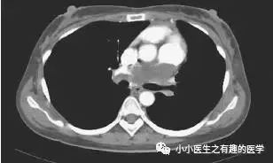
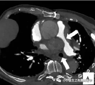
法兰西报道的一个患者。
这么大一个血栓!
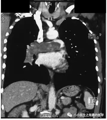
好大的一条血栓!
然而,最终做了手术,病理如下:
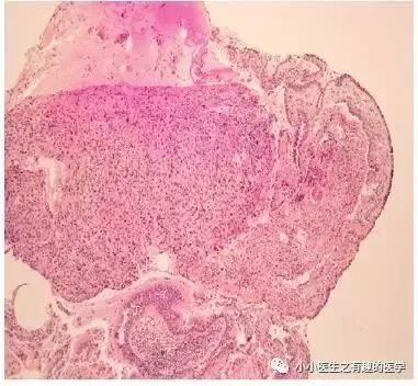
五颜六色的,好好看啊。然而,this is 肺动脉内膜肉瘤。
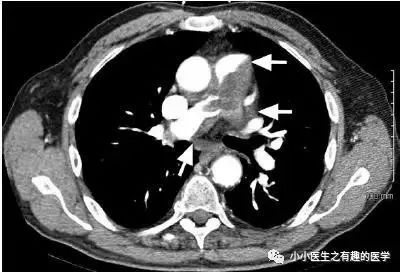
仔细看看,不对。
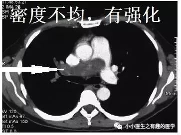
充盈缺损的地方,密度不均匀,并且有强化,血栓里面没有血管,一般不会强化,所以有可能是肿瘤。
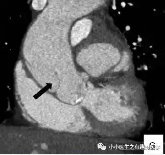
足球王国巴西报道的一例。
这是什么?
病理:Intimal sarcoma of the pulmonary artery,
肺动脉内膜肉瘤
。
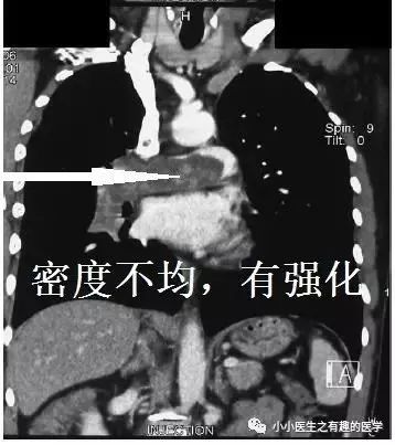
仔细看看:
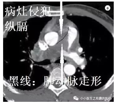
CT示肺动脉内肿块,肿块侵犯纵隔,血栓不会跑到
纵隔
。
德国蒂宾根大学。
女性,33岁,反复劳累后呼吸困难5月,胸膜炎性胸痛,伴有咳嗽,以肺栓塞可能收入院。
A 33-year-old female with a 5 month history of recurrent episodes of exertional dyspnoea, pleuritic chest pain and an urge to cough was referred to hospital with the tentative diagnosis of pulmonary embolism.

看CT,左肺动脉血栓了。
患者结局呢?

遗体解剖发现肺动脉肿块,延伸至右心房。病理:
原发性肺动脉绒毛膜癌
。
At autopsy, a tumour was found occluding the left pulmonary artery extending into the right atrium. Histological evaluation showed a primary choriocarcinoma.
这不是癌栓,这是肺动脉的肿瘤。
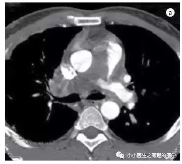
44岁女性,平素体健,进行性的呼吸困难3月,双侧踝周水肿。
A 44 year old woman, who had enjoyed good health up until recently, presented with a three month history of progressive shortness of breath and bilateral ankle oedema.
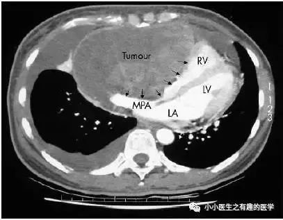
LV:左心室,不是LV包包。MPA:主肺动脉。
这个相对简单,是肺动脉严重受压了,引起类似充盈缺损的改变。
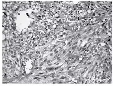
纵隔镜检查,病理:
恶性梭形细胞肿瘤
。
Mediastinoscopy and biopsy of the tumour was subsequently performed which yielded malignant spindle cell tumour by histology.
82岁女老人,劳力性呼吸困难,近期加重。
An 82-year-old woman with no previous history of heart or lung diseases presented with exertional dyspnoea and episodes of an acute shortness of breath over the preceding month.
看CT,妥妥的血栓了。
疑诊肺栓塞,抗凝治疗。
The patient was initially anticoagulated for presumed pulmonary thromboembolism.
患者血流动力学恶化,心源性休克死亡。
The patient suffered haemodynamic deterioration and died from cardiogenic shock.
为什么涅?为什么按照肺栓塞治疗,患者还是去见马克思了?偶,歪国人喜欢见上帝。
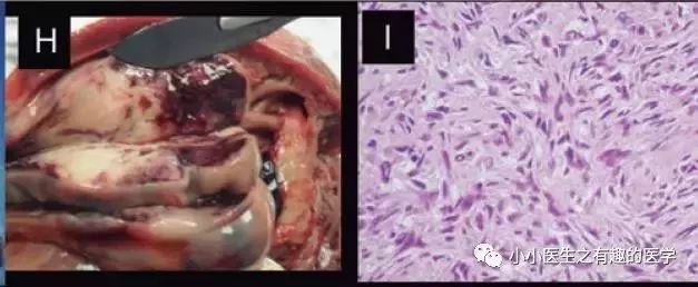
肿么办?你的灵魂去见上帝,你的肉体给我解剖学习。
病理:
肺动脉内膜肉瘤
。
Macroscopic (Panels G and H) and microscopic (Panel I) examination revealed the presence of an intimal sarcoma of the pulmonary artery.
里约热内卢联邦大学。
35岁女性,呼吸困难,咯血,胸痛。哈哈,典型的肺栓塞。
绝对的肺栓塞。
诊断慢性肺栓塞,治疗6月。
复查:
星星,还是那颗星;月亮,还是那个月......,
血栓,还是那么大滴哟,一个——血——栓。还来不及手术,患者died。
A significant clinical worsening was observed and the patient died before she could be submitted to a diagnostic/therapeutic surgical procedure.
最终诊断:
肺动脉肉瘤
。
美国德克萨斯州心脏研究所。
77岁男性,外院转入。
A 77-year-old man was transferred from another hospital for treatment of a saddle PA embolus.
He had been treated with thrombolytic and anticoagulant agents without clinical improvement.
然而,这个血栓怎么看着乱七八糟的?这是一个不正经的血栓。怎么办?牛叉的医院就喜欢简单、粗暴:
切开肺动脉见肿块。
We incised the main PA and inspected the mass.
肺动脉病理:
高分化肉瘤
。
The histopathologic report showed a high-grade sarcoma
with focal myogenic and chondrogenic differentiation.
小Japan的报道。39岁女性,呼吸困难。
A 39-year-old woman with shortness of breath was admitted to our hospital on October, 2009
.
不可否认,这个血栓很温柔,很典型,很丝滑。
诊断肺栓塞,抗凝治疗。
On admission, we diagnosed the patient to have a pulmonary thromboembolism and initiated anti-thrombotic therapy with alteplase at 24,000,000 U div for one hour followed by anti-coagulant therapy with heparin at 15,000 U/day.
然后,患者感染,心衰,就给予美罗培南抗感染
(真高级,日本也喜欢高级抗生素)
,多巴胺强心。
Day 11, the patient developed heart failure due to infection and treatment with an antibiotic (MEPM 1 g/day) and a catecholamine (Dopamine) was started.
然而,抗凝无效。
术后病理:
肺动脉内膜肉瘤
。
Histopathologically, the surgical specimen was not found to be thrombotic tissue but rather an intimal sarcoma of the pulmonary artery.
意大利University of Milan。患者84岁,呼吸困难,胸痛。
We report the case of a 84-year-old patient, with a history of coronary artery bypass graft。The patient referred to our emergency department for chest pain and dyspnoea.
CT提示肺动脉受压
(纵隔血肿压迫)
。进一步检查发现A型主动脉夹层。
CT pulmonary angiography did not show PE but a significant pulmonary artery compression。A subsequent arterial phase,
demonstrated a TypeAaortic dissection.
结局:The patient died 3 days after CT examination.
71岁男性,诉胸骨后胸痛。
A 71-year-old man was admitted owing to an episode of retrosternal chest pain.
肺通气灌注扫描:
右肺大面积肺栓塞
。
Pulmonary perfusion scintigraphy showed the total absence of perfusion in the right lung, consistent with massive pulmonary embolism in the right pulmonary artery.
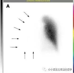
下一步肿么办?抗凝?NO。歪国人的处理是:
肺动脉CTA
。结果:
主动脉夹层,木有血栓
。
To obtain a definite diagnosis, chest computed tomography was carried out, which, unexpectedly, disclosed a type II
dissecting aneurysm of the ascending aorta.


