

鉴于很多读者的建议,丁香园
肿瘤时间(oncolatdxy)
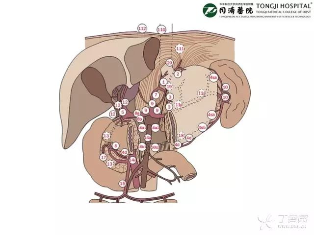
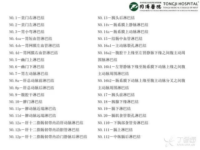
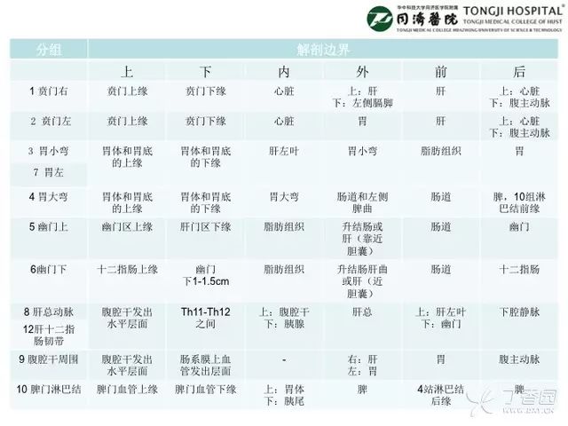
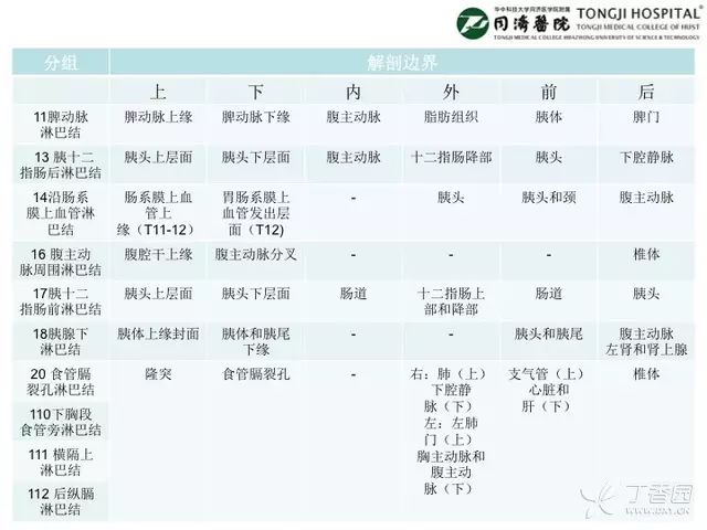
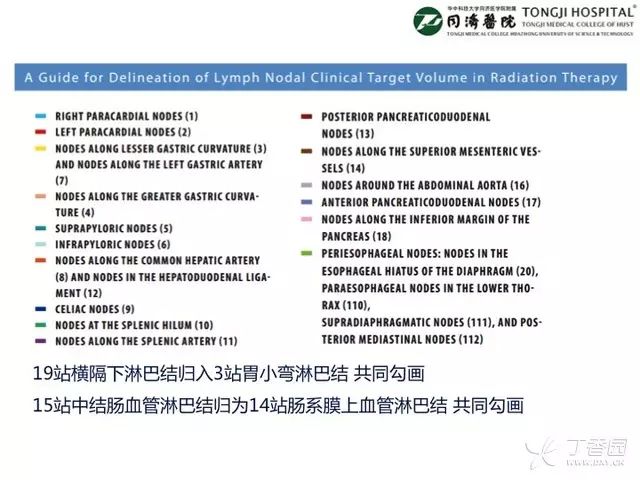
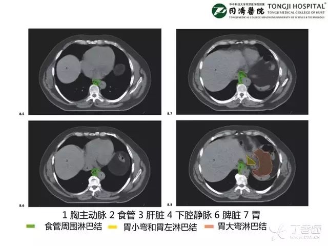
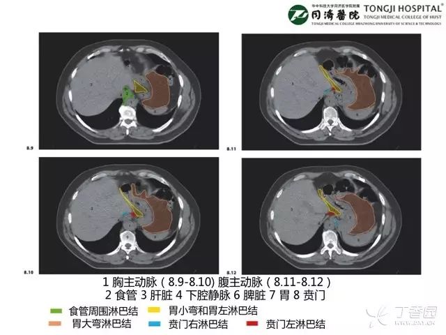
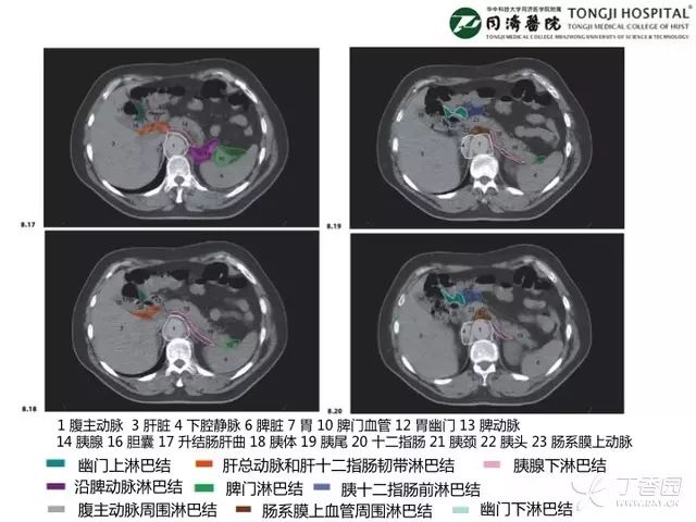
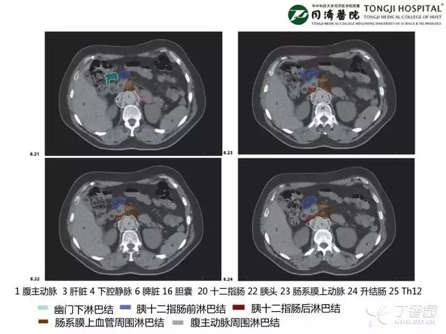
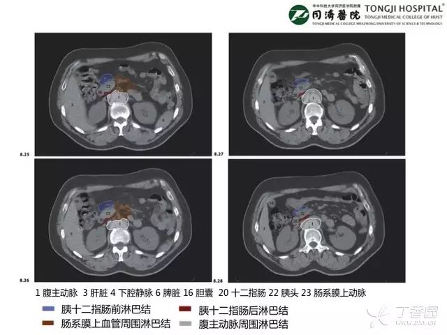
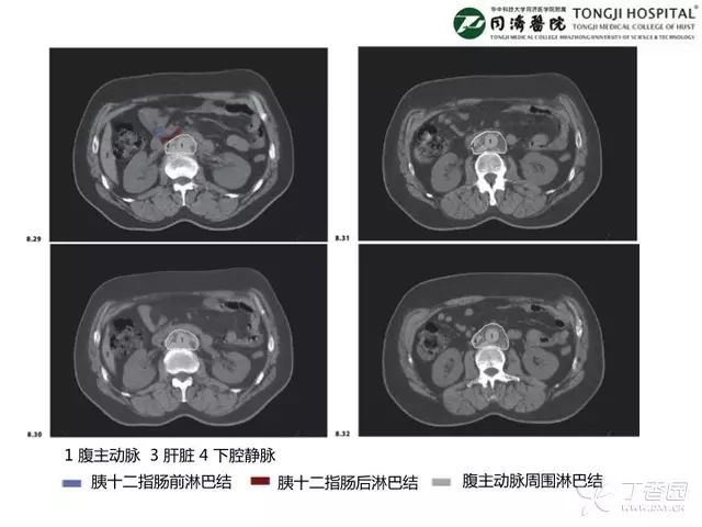
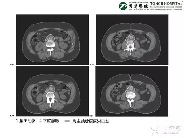
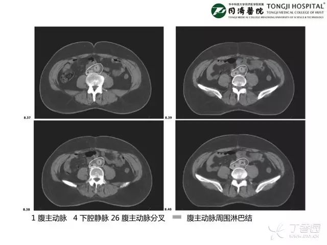
现推出上腹部淋巴结引流区图谱,希望能够帮助到大家。
如果你对上腹部解剖影像还没有完全掌握,建议先看几遍
掌握这篇,晋级腹部 CT 影像高手
,不然看完这篇可能会一脸懵逼。对于上腹部解剖影像,你需要掌握几个关键点:1. 肝门结构;2. 肠系膜上动、静脉的走形;3. 十二指肠走形;4. 腹腔干分支走形;5. 门静脉的走形和伴行结构。
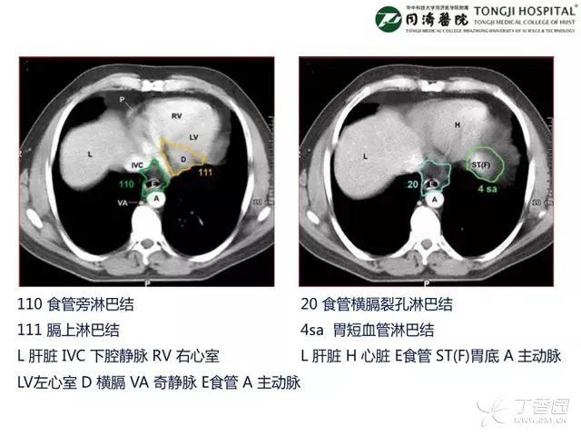
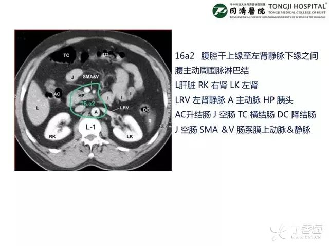
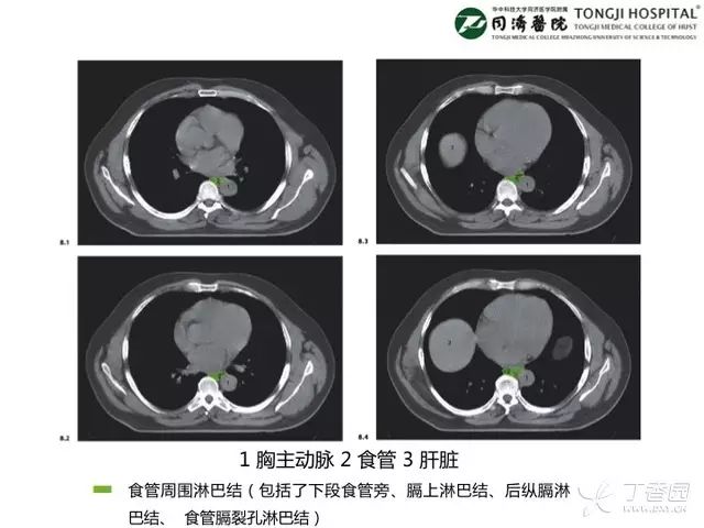
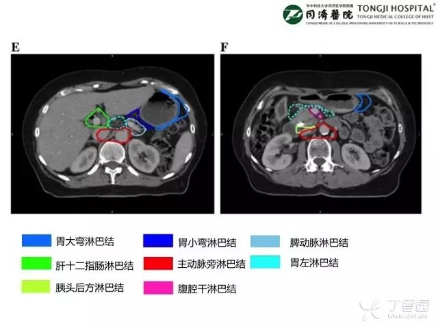
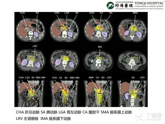
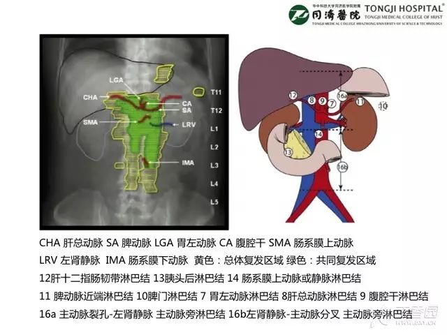
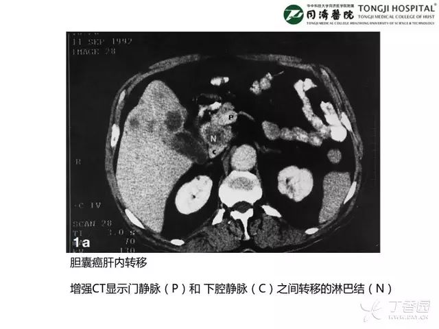
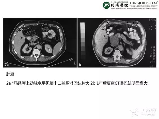
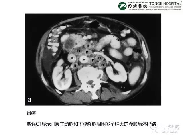
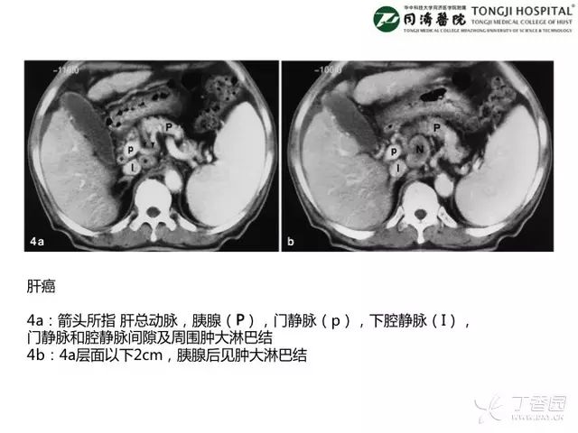
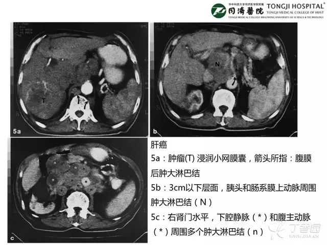
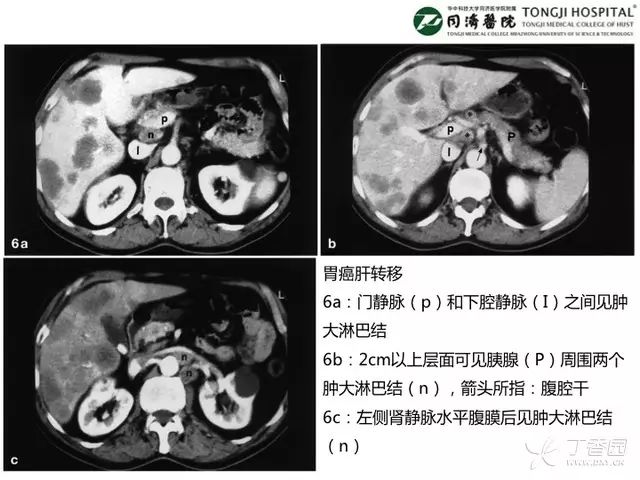
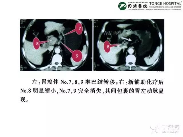
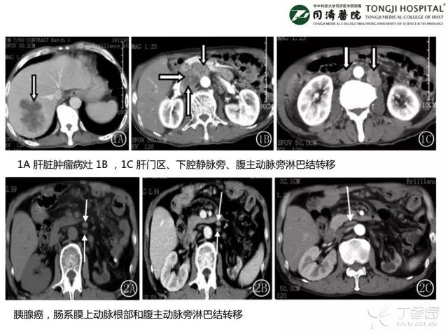
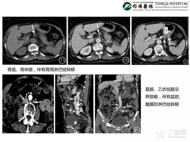
为了让大家看清上腹部淋巴结转移是什么样子滴,文末附赠几张已经在期刊发表的 CT 影像。


如果这个 PPT 对你有用,欢迎转发给身边的小伙伴,大家一起涨姿势。
本文作者:华中科技大学同济医学院附属同济医院肿瘤中心 刘东伯
来源:微信公众号 dongboliu251
编辑:汪小鱼 | 题图来源:视觉中国


参考文献:
1. 李国立. CT分组定位诊断法在胃癌淋巴结转移中的临床应用. 消化肿瘤杂志(电子版)2009 年 9 月第 1 卷第 1 期。
2. Jennifer Y
et al
.
Gastric lymph node contouring atlas: A tool to aid in
clinical target volume definition in 3-dimensional
treatment planning for gastric cancer. Practical Radiation Oncology (2013) 3, e11–e19.
3. 叶小青等. 多层螺旋 C T 在消化系统常见肿瘤淋巴结转移诊断中的临床价值. 医学影像学杂志 2015 年第 25 卷第 4 期.
4. S. C. Efremidis
et al
. Pathways of lymph node involvement in upper abdominal malignancies: evaluation with high-resolution CT. Eur. Radiol. 9, 868±874 (1999).
5. Oscar Matzinger
et al
.
EORTC-ROG expert opinion: Radiotherapy volume and treatment guidelines
for neoadjuvant radiation of adenocarcinomas of the gastroesophageal junction and the stomach. Radiotherapy and Oncology 92 (2009) 164–175.
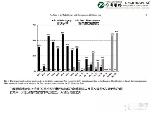
6. Hong In Yoon
et al
. Defining the target volume for post-operative radiotherapy after D2 dissection in gastric cancer by CT-based vessel-guided delineation. Radiother Oncol. 2013 Jul;108(1):72-7.
7. GA Cefaro
et al
. A Guide for Delineation of Lymph Nodal Clinical Target Volume in Radiation Therapy. Springer Berlin Heidelberg , 2008.


