BACKGROUND: Neuronavigation is an established technology in neurosurgery. In parts of the world and certain circumstances in which neuronavigation is not easily available or affordable, alternative techniques may be considered.
背景:神经导航技术在神经外科已经很成熟,但是,世界的某些地区或在某种特殊情况下,神经导航无法实施或无法负担时,可考虑神经导航的替代技术。
OBJECTIVE: An app to assist scalp localization of superficial supratentorial lesions has been introduced, and its accuracy has been compared with established neuro-navigation systems.
目的:一款用于辅助幕上浅表病灶头皮定位的手机App已经上市,将其精准度与已经很成熟的神经导航系统进行比较。
METHODS: Sina is a simple smartphone app that over-laps the transparent patients’ computed tomography/mag-netic resonance images on the background camera. How to use Sina intraoperatively is described. The app was used for scalp localization of the center of the lesions in 11 patients with supratentorial pathologies <3 cm in longest diameter and <2 cm from the cortex. After localization of the lesion using Sina, the center of the lesion was marked on the scalp using standard neuronavigation systems and the deviations were measured.
方法:Sina是一款简单的智能手机App,可将患者透明的CT或MRI影像与相机背景进行重叠。文中将描述如何在术中使用Sina。该App对11例患者(幕上病灶的最大直径<3cm且病灶距皮层<2cm)的病灶中心进行头皮定位。然后与标准神经导航系统在头皮标记的病灶中心进行比较,并测量两者误差。
RESULTS: Implementation of Sina for intraoperative scalp localization is simple and practical. The center of the lesions localized by Sina was 10.2±2 mm different from localization done by standard neuronavigation systems.
结果:Sina用于术中头皮定位简单实用。与标准神经导航系统定位的病灶中心的相比,两者测量结果相差10.2±2mm。
CONCLUSION: When neuronavigation is not easily available or affordable, Sina can be helpful for scalp localization and preoperative planning of the incision for selected supratentorial pathologies.
结论:如果神经导航无法实施或不能负担时,Sina可辅助合适的幕上病灶进行头皮定位和规划术前切口。
INTRODUCTION 前言
Neurosurgeons and neurosurgical trainees, over time, learn how to correlate image studies with patient anatomy and localize the lesions in the nervous system.
时光流逝,神经外科医生和准神经外科医生正不断学习如何将患者的影像结果与实际解剖结合起来,进行神经系统病灶的定位。
However, that is not accurate enough and neuronavigation has become an established technology in neurosurgery. In certain circumstances and in parts of the world, neuronavigation is not easily available or affordable.
虽然并不完全准确,但神经导航逐渐成为神经外科的成熟技术。在某些情况下,某些地区,神经导航无法实施或无法负担。
Popularity and availability of smart devices including smart phones make them a very good platform for augmented reality purposes and intraoperative localization. There are available apps that could be used for the same purposes; however, the need for certain features and potential for further development prompted the author to consider developing a simple app for scalp localization and intraoperative planning.
包括智能手机在内的智能设备的普及和实用,使他们成为一个很好的平台,可以实现增强现实的目的和术中定位。有很多诸如此类的软件,但是某些特定需要和进一步发展的潜在需求促使笔者去研发一款简单实用的App,以进行术前头皮定位和规划切口。
In this study the author introduces Sina Intraoperative Neurosurgical Assist (Sina) as a simple app to assist image-based scalp localization. It is an Android app written in Java available for download from Google Play Store.
在本研究中,笔者将介绍一款基于影像辅助头皮定位的简单App,Sina神经外科术中助手(Sina)。Sina是用Java编写的Android App,可从Google Play商店下载。
In order to investigate the functionality of Sina in circumstances in which neuronavigation is not available, the accuracy of Sina has been compared with the established neuronavigation systems.
为了研究神经导航系统无法实施时Sina的性能,将其精准度与成熟的神经导航系统进行比较。
METHODS 方法
App Instructions App使用说明
Sina simply overlaps the transparent patients’ computed tomography/magnetic resonance imaging (CT/MRI) on the background camera. The first screen of the app shows the instructions for use. There is a “SELECT IMAGE” button for selection and loading the images from gallery of the android device. After selection, the image will be transparent and overlapped on the camera. While moving the device toward or away from the patient’s head, the image can be overlapped properly. Magnification cannot be modified through Sina.
Sina可简单的将患者透明的计算机断层扫描/核磁共振成像(CT / MRI)重叠到
相机背景上。
该App的第一个界面为使用说明。
有一个“SELECT IMAGE(选择图像)”按钮,用于选择和加载Android设备库的图像(CT/MRI)
。之后,图像将变成透明的,并重叠在相机背景上。
移动相机靠近或远离病人的头部时,图像可与患者头部恰当的重叠。
Sina不具有缩放功能。
Because CT/MRI convention is as if one is viewing from below rather than above the head, the orientation of the loaded image is usually different from the patient’s position. Therefore double tapping the image with Sina to flip the image is helpful.
因为CT或MRI的片子中“左右”是反着的,所以,使用Sina时可以双击屏幕,将半透明化的影像左右对调,有助于操作。
平台审稿团汤可博士翻译:由于CT/MRI的断层图像通常为足侧观,在患者的头侧定位加载影像时常常发生位置反向,因此使用Sina时可以双击翻转图像来进行纠正!
Intraoperative Technique 术中操作
Appropriate axial, coronal, and sagittal CT or MRI images best showing the lesion are taken by an Android device camera in portrait mode. These images could be taken by other cameras and transferred to the Android device with or without editing, before use by Sina.
用Android相机以人像模式拍摄可最佳显示病灶的轴位,冠状位和矢状位CT或MRI图像。
这些图像也可以用其他相机拍摄,无论是否进行编辑,在使用Sina之前传输到Android设备上。
After anesthesia and before positioning (while the patient is in neutral supine position), the coronal suture is localized on the skull and the midline of the head is marked using anatomic landmarks. Sina is launched, and the axial image is loaded as the first step. The side of the lesion is verified as a routine part of the time-out procedure. An assistant puts her or his finger on coronal suture so that the tip of the finger can be used as a reference point for contour of the skull. Using coronal suture and midline as anatomic guides, the axial image is overlapped on the head from the top of the patient so that the coronal suture of the head matches with its location on the image. Localizing the center of the lesion, a coronal line is drawn on the head by the assistant (Figure 1). Any other landmarks, like a skull dent, or radiodense markers that can be matched with its location on the images can also be used.
麻醉后,摆体位前(患者处于正中仰卧位),使用解剖标志在颅骨上画出冠状缝,并标出中线的位置。
打开Sina,第一步先加载轴位图像。确认病灶侧。助手将其手指放在冠状缝上,可把指尖看作是颅骨轮廓的参照点。
把冠状缝和中线作为解剖标志,将轴位图像在患者头顶重叠,使冠状缝与其在图像上的位置吻合。
定位病灶中心时,助手在患者头部画出冠状线(图1)。也可以使用其他解剖标志,如颅骨凹痕或可匹配其在图像上位置的不透X线的标记物。

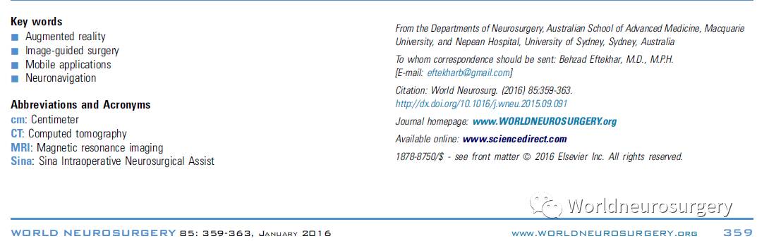
Figure 1. This is a reconstructed image (not taken intraoperatively) showing an example of overlapping of the axial image and drawing of the coronal line accordingly. On the right, localizing the center of the lesion, a coronal line is drawn on the head
图1.这是一个重建图像(非术中拍摄),为轴位图像与在头颅上绘制的相应的冠状线重叠的示例。为定位病灶中心,在右图的头上画出冠状线。
The coronal image is loaded as the next step. The assistant puts a finger on the coronal line that was drawn in the previous step so that the fingertip can be used as a reference point for contour of the skull at the coronal plane of the lesion. Using the midline and tip of the finger as guides, the coronal image is overlapped on the head. In more anterior lesions, the nose and orbital rim can also be used as guides to overlap the images over the head. After properly localizing the center of the lesion, an anteroposterior line is drawn by the assistant (Figure 2). The room lights may interfere with using the device and may need to be temporarily adjusted.
第二步是加载冠状位图像。助手将手指放在上一步画出的冠状线上,使指尖能够用作病灶在颅骨轮廓冠状平面上的参照标志。使用中线和指尖作为参照,将冠状位图像在头部重叠。更靠前的病灶,鼻子和眼眶也可以用作在头部重叠图像的参照。
准确定位病灶中心之后,由助手画出病灶的前后线(图2)。如果房间的灯光干扰设备的使用,可临时调整。
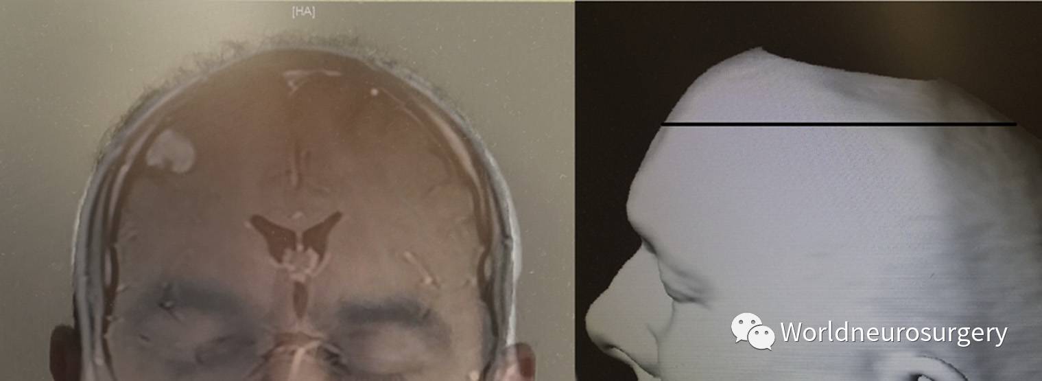
Figure 2. This is a reconstructed image (not taken intraoperatively) showing an example of overlapping the coronal image on the head, so that the contour of the head and midline match with the image. On the right, localizing the center of the lesion, an anteroposterior line is drawn on the head perpendicular to the scan.
图2.这是一个重建的图像(非术中拍摄),为冠状位图像在头部重叠的示例,使头部轮廓和中线与图像吻合。为定位的病灶中心,在右图头部垂直扫描方向画出前后线。
The crossing point between two drawn lines is considered the estimated center of the lesion (Figure 3). Optionally the sagittal cut can be loaded to check the accuracy of previous localization anteroposteriorly. Again, coronal suture should be used as the guide. For lesions close to the midline, using the sagittal image first and coronal image second works better.
两条线的交点是估算的病灶中心(图3)。选择加载合适的矢状位切面以检验之前前后方向上定位的准确度。再次把冠状缝作为参照。对于靠近中线的病灶,先用矢状位图像,再用冠状位图像较好。
Comparing the Accuracy of Sina with Standard Neuronavigation Systems
Sina与标准的神经导航系统精准度的比较
Because the accuracy of scalp incision matters more when the neurosurgeon deals with small lesions, the accuracy of Sina was compared with known neuronavigation systems for small superficial lesions. Lesions <3 cm in longest diameter were arbitrarily defined as small.
在处理小的病灶时,头皮切口位置的准确性非常重要,所以我们将Sina对小的表浅病灶的精准度与我们熟知的神
经导航系统相比较。
我们把最长径<3cm的病灶定义为小病灶。
In 11 patients with supratentorial lesions <3 cm in longest diameter and <2 cm deep from the cortex, using the previously mentioned technique on Galaxy Note 3 5.700, the lesions were localized by the author. After localization of the lesion using Sina, the center of the lesion was localized and marked (by an assisting registrar or fellow), using standard neuronavigation systems (one case StealthStation, Medtronic, Minneapolis, Minnesota, USA, and the rest Brainlab, Munich, Bavaria, Germany). A ruler was placed on the site for measurements.
11例幕上病灶最长径<3cm,距皮层<2cm的患者,笔者在Galaxy Note 3 5.700上使用前面提到的方法定位病灶。使用Sina定位病灶后,再使用标准的神经导航系统(1例
使用StealthStation导航系统(Medtronic, Minneapolis, Minnesota, USA),其他的使用Brainlab导航系统(Munich, Bavaria, Germany))定位,并在住院医师或同事的帮助下标出病灶中心。用尺子测量两种方法定位的病灶中心的差值。
The deviation of alignment (mm) was calculated by dividing the distance (pixels) between the localizations (A) by length (pixels) of 1 cm cut of ruler (CM) times 10 (10A/CM) (Figure 3). In order to reduce the error due to manual intervention, each measurement was done twice and then averaged. The deviations (mm) and other variables including age/sex, site, and size of the lesion and pathology were tabulated. All initial measurements were made in pixels using the computer software Paint (v6.2 Microsoft, Redmond, Washington, USA). The mean and standard deviation of the differences and durations were calculated using computer software Calc (LibreOffice 4.2.5.2, The Document Foundation, Paris, France).
两定位点(A)间的距离(像素),即校准偏差(mm),通过分成10份(10A/CM)的1cm的尺子来测量计算(图3)。 为了减少人为干扰的误差,每次测量两次,然后取平均值。列出偏差(mm)和包括年龄/性别,病灶的部位和大小,以及病理类型的其他变量。所有初始测量结果均使用计算机软件Paint(v6.2 Microsoft,Redmond,Washington,USA)进行运算。 使用计算机软件Calc(LibreOffice 4.2.5.2,The Document Foundation,Paris,France)计算差值和定位所耗费时间的平均值和标准差。
Figure 3. The image was taken intraoperatively after localizations done by smartphone app to assist scalp localization of cranial lesions is introduced. Sina (blue lines) and neuronavigation systems (black dot). The deviation of alignment (A) and 1 cm length of the ruler (CM) are marked.
图3.智能手机App辅助完成颅内病灶头皮定位术中拍摄的图片。Sina的定位(蓝线)和神经导航系统的定位(黑点)。 标记的校准偏差(A)和1cm长的尺子(CM)。
RESULTS 结果
Localization using Sina added on average 4±1 minutes to the operating time. Taking photos of the patients’ images were not included in the timing. Considering the fact that the prerelease versions of the app had been used by the author during app development, no comment could be made on the learning curve.
使用Sina定位操作时间平均增加4±1min。拍摄患者影像图片时间不包含在内。 考虑到在App更新过程中笔者已经使用过试用版,故对学习曲线不做评论。
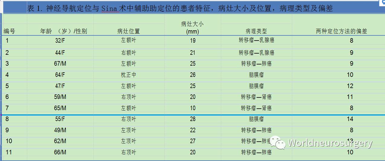
Table 1 shows the size of the lesions, pathology, and difference between neuronavigation and localization by Sina. The mean difference is 10.2±2 mm with an 8- to 14-mm range.
表1显示了病灶的大小,病理类型,以及神经导航定位和Sina定位的差异。两者之间平均差为10.2±2mm,相差范围为8〜14mm。
Stealth was used for the second patient. Her lesion was 2 cm deep to the cortex. Brainlab was the neuronavigation system for the rest of the patients.
The directions of misalignments did not follow any specific pattern (was not predominantly craniocaudal or anteroposterior).
第二个病人用了Stealth导航系统。她的病灶距皮层2cm深。其余的病人使用Brainlab导航系统。
方向的偏差不遵循任何特定的模式(主要不是头尾位或前后位)。
What Is the Likely Error If the Scan Has Not Been Acquired in Orbitomeatal Plane or Is Tilted?
如果影像没有按照眶耳平面扫描或有所倾斜,头皮定位时可能出现什么样的误差呢?
Considering the fact that the rotation may happen around different axes and also that head shapes and location of the lesions are different, coming up with a generalized formula may not be practical.
考虑到旋转可能发生在不同轴位周围,且头形和病灶位置也不尽相同,推出一个广泛适用的公式也不切实际。
However, in order to estimate the error, the author has assumed rotation of the head around the line that joins 2 external auditory meatuses, in a special case of a lesion in the midline vertex. Figure 4 shows that the targeting anteroposterior error (mm) is estimated by tangent of tilting angle multiplied by the auricular height. The auricular height varies in different races; however, for a height of 120 mm, 1 degree of tilt can cause around 2 mm of targeting deviation.
但是,为了估算误差,在一例病灶位于中线顶点的特殊病例中,笔者假定头部以两外耳道连线为轴旋转。 图4显示,通过倾斜角的正切值乘以耳廓高度来估算定位点前后位误差(mm)。
耳廓高度与种族有关; 但是,对于120mm的高度,1度的倾斜可导致约2mm的定位偏差。
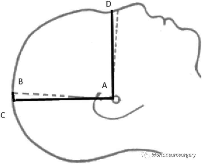
Figure 4. The scan should be acquired with AD as the orbitomeatal plane and AC as the auricular height. If we assume that the scan has been tilted for an angle (∠CAB in our special case), the center of the lesion is calculated as B instead of C (with CB as targeting error). The targeting anteroposterior error (CB) can be estimated by tangent of tilting angle
(:CAB) multiplied by the auricular height (AC). Due to the contour and shape of the head, the calculation is only an estimate for practical purposes and becomes less accurate as the angle increases. A smartphone app to assist scalp localization of cranial lesions is introduced.
图4.图像扫描本应获得如AD所示的眶耳平面,和AC所示的耳廓高度。 我们假设扫描已经倾斜了一个角度(我们这例特殊病例中为∠CAB),病灶的中心应以B代替C来计算(CB为误差)。 前后位误差(CB)可用倾斜角(∠CAB)的正切值乘以耳廓高度(AC)来估算。 由于头部的轮廓和形状的差异,计算结果仅是粗略估算值,随着倾斜角度的增大,准确性逐渐下降。 于是引入一款智能手机App辅助颅内病灶的头皮定位。
In practice, the majority of lesions are not in the midline and may be located at different heights from the orbitomeatal plane. Assuming the average height of the lesions from the orbitomeatal plane to be 60 mm (half of the auricular height), 1-mm targeting deviation for 1 degree of tilt seems to be a better estimate of the average error for practical purposes. It should be taken into consideration the presumption about the shape of the head and the fact that due to contour of the head, with increase of the tilting angle the estimated error using the abovementioned formula becomes less accurate.
实际情况中,大多数病灶不在中线,距眶耳平面的高度也不尽相同。假设病灶距眶耳平面的平均高度为60mm(耳廓高度的一半),
1°倾斜的1mm偏差是对实际病灶的平均误差的更好的评估。
应考虑到头形的问题,以及由于头部轮廓的问题,随着倾斜角度增大,使用上述公式评估误差的准确度下降。
DISCUSSION 讨论


